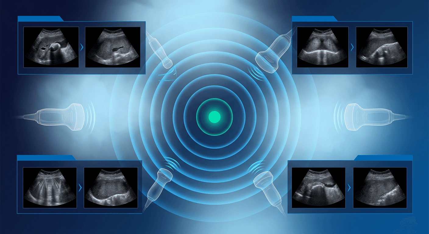
Characterizing renal cysts ultrasound findings starts with a binary decision: does the lesion meet strict simple-cyst criteria, or do wall, septal, or solid features require further imaging? Practicing sonologists who apply those criteria consistently can leave most incidental cysts alone, escalate the right cases, and avoid both over-calling debris and missing a cystic neoplasm. Ultrasound is often the first test that shows a kidney cyst, whether the study is dedicated renal imaging or part of a broader abdominal survey. Size alone does not tell you whether a cyst is dangerous. Morphology, Doppler, and comparison with prior studies do.
Renal Cysts Ultrasound: Applying Simple-Cyst Criteria
Call a lesion a simple cyst only when every criterion is met after the image has been optimized:
- Anechoic contents, with no persistent internal echoes
- Thin, smooth, imperceptible wall
- Round or oval shape and a sharp parenchymal interface
- Posterior acoustic enhancement
- No septa, mural nodules, or calcifications
- No internal color or spectral Doppler flow
If any item fails, do not use the phrase simple cyst. Name the feature that broke the rules and recommend the next step.
Call a cyst simple only when it is anechoic, thin-walled, and free of septa, nodules, and Doppler flow after technical optimization.
Before you decide, reduce gain, use harmonic imaging, scan the entire wall in two planes, and interrogate with color Doppler sensitive to low flow. Superficial cysts in thinner patients often look cleaner with a higher-frequency or linear transducer. Measure the largest diameter and document laterality and pole. These habits fit the same systematic renal sweeps used in a complete abdominal ultrasound protocol.
Features That Make an Ultrasound Renal Cyst Complex
Complexity is a continuum. Describe each finding rather than assigning a Bosniak number that gray-scale ultrasound cannot fully support.
Septa and wall
A few thin, smooth septa may still represent a minimally complex cyst. Thick, irregular, or numerous septa, or septa with detectable flow, raise concern and should go to CT or MRI. Focal or diffuse wall thickening and an irregular contour likewise take the lesion out of the simple category.
Solid components, calcification, and internal echoes
Any mural nodule or papillary projection needs Doppler interrogation. Flow within a nodule is a red flag for a cystic neoplasm. Debris, clot, or milk of calcium can mimic a nodule; change the patient's position and rescan. Thin, smooth calcification can be benign. Thick or irregular calcification is better characterized on CT. Low-level internal echoes may represent hemorrhage, proteinaceous content, infection, or artifact. A hemorrhagic cyst can look complex on ultrasound and still prove benign on CT or MRI. Do not assume malignancy, and do not assume benignity. Recommend contrast-enhanced CT or MRI when echoes persist after optimization.
Wall thickening, mobile debris, and surrounding inflammatory change in a febrile patient may indicate an infected cyst or abscess. Correlate with the clinical picture. Imaging does not replace that assessment.
Bosniak Features and How SRU-Style Ultrasound Guidance Differs
The Bosniak classification was built for CT and is widely applied to MRI. It groups cystic renal masses by wall and septal thickness, number of septa, calcification, and enhancement of solid elements, from simple (I) through minimally complex (II), follow-up lesions (IIF), surgically complex (III), and frankly malignant-appearing cystic masses (IV).
Ultrasound can show many of those morphologic features, but it does not demonstrate enhancement the way multiphase CT or MRI does. That limitation matters. A confident ultrasound diagnosis of a simple cyst corresponds to Bosniak I and, in ordinary practice, needs no follow-up. Mild complexity may correspond to Bosniak II or IIF, yet IIF, III, and IV should generally be assigned on CT or MRI unless your laboratory uses contrast-enhanced ultrasound with defined local protocols.
SRU consensus guidance on incidental cystic renal masses is more ultrasound-centric than classic Bosniak numbering:
- Strictly simple cysts, including large ones, typically need no imaging follow-up
- Minimally complex cysts with thin septa or thin calcification may be left alone or sent for characterization, depending on the exact features and the patient
- Nodularity, thick septa, or solid elements should go to CT or MRI rather than ultrasound follow-up alone
When the two frameworks diverge, say so. It is fair to report that a lesion meets ultrasound criteria for a simple cyst (Bosniak I equivalent) or that it is complex and requires contrast-enhanced CT or MRI for Bosniak classification. It is not fair to assign Bosniak III or IV from gray-scale ultrasound alone. A younger adult, a hereditary renal cancer syndrome, or a lesion that is growing or newly complex deserves a lower threshold for CT or MRI even if the ultrasound appearance is only mildly imperfect.
Mimics, Artifacts, and Pitfalls
Exclude look-alikes before you commit to a cystic mass.
- Localized hydronephrosis or a calyceal diverticulum can mimic a cyst. Look for communication with the collecting system and for dependent milk of calcium.
- Parapelvic cysts occupy the sinus and can be mistaken for hydronephrosis. They do not connect to calyces, and the collecting system remains decompressed.
- Anechoic hilar vessels, including a rare aneurysm, show flow. Doppler every apparently cystic structure in the hilum.
- Reverberation and slice-thickness artifact create false echoes or a pseudo-septum. Change the angle, narrow the field, and use harmonics.
- Hemorrhagic or proteinaceous cysts look complex on ultrasound and still may be benign after CT or MRI.
- Cystic renal cell carcinoma and multilocular cystic neoplasms can be relatively paucicellular. Any mural nodule or thick septum is enough to stop calling the lesion simple.
- A few age-related simple cysts are not the same diagnosis as polycystic kidney disease. Number, distribution, and clinical context belong in the interpretation, but the ultrasound appearance alone does not establish a hereditary cystic disease.
In the renal transplant, cysts may involve the allograft or the native kidneys. Dedicated transplant scanning answers vascular and collecting-system questions that incidental cyst work does not replace. See ultrasound evaluation of the renal transplant for that protocol.
When Ultrasound Is Enough and When CT or MRI Is Needed
Ultrasound is enough when every simple-cyst criterion is met, the finding is incidental, and there are no high-risk clinical features. You do not need to send every cyst to CT.
Recommend CT or MRI when:
- Simple-cyst criteria fail after optimization
- There is a mural nodule, thick septum, or internal Doppler flow
- The lesion is incompletely seen because of body habitus, gas, or location
- There is unexplained hematuria, a suspected solid mass, or a hereditary renal tumor syndrome
- Priors show growth of solid elements or new complexity
- The question is infection, trauma, or a symptomatic cyst under consideration for intervention
Contrast-enhanced ultrasound can show enhancing septa or nodules and is used in some centers when iodinated contrast or gadolinium is undesirable. Availability varies. Gray-scale ultrasound does not substitute for contrast assessment of complex lesions.
Size, symptoms, and treatment
There is no size cutoff that makes a simple cyst cancerous. A small cystic mass with a nodule is more concerning than a large simple cyst that meets every criterion. What counts as a large kidney cyst is not standardized. Some practices comment when a cyst measures several centimeters because of mass effect, pain, or collecting-system compression. Aspiration or unroofing is a clinical decision based on symptoms, obstruction, or infection, not on diameter alone. Ultrasound documents size, location, and effect on the collecting system. It does not decide whether a given patient needs treatment.
Reporting Pearls
- Write simple cyst only when all criteria are documented
- Give laterality, pole, cortical versus parapelvic location, and maximum diameter
- For multiple cysts, report the largest and any that are complex
- Describe complexity in plain language: septa, wall, nodules, calcification, Doppler flow
- Do not assign a numeric Bosniak category from noncontrast ultrasound
- Recommend no follow-up for strictly simple incidental cysts, or CT or MRI for incomplete or complex lesions, consistent with current SRU-style and institutional guidance
- Compare with priors and state whether the finding is new, stable, or changed
A disciplined ultrasound renal cyst assessment is mostly about saying simple when it is simple and stopping when it is not. Optimize the image, apply strict criteria, Doppler anything that looks solid, and send complex or incompletely seen lesions to CT or MRI for true Bosniak classification. Size is a measurement, not a malignancy score. IAME offers accredited ultrasound CME courses in abdominal imaging for clinicians who want structured review of cystic renal and other abdominal findings.
Frequently Asked Questions
What are the ultrasound classifications of renal cysts?
Ultrasound does not replace the CT and MRI Bosniak system, but it does separate simple cysts from complex cystic masses. Simple cysts are anechoic, thin-walled, and without septa, nodules, or flow. Complex cysts are described by their septa, wall, calcification, and solid elements, then referred for contrast-enhanced CT or MRI when those features are present.
What test shows kidney cysts?
Ultrasound, CT, and MRI all show kidney cysts. Ultrasound is often the first test and is sufficient when a cyst meets strict simple-cyst criteria. CT or MRI is used when the cyst is complex, incompletely characterized, or when enhancement of septa or nodules must be assessed.
How to determine if a kidney cyst is cancerous?
No single ultrasound feature proves cancer, and a lesion that meets simple-cyst criteria should not be called malignant. Suspicion rises with Doppler-positive nodules, thick irregular walls or septa, and solid components. Risk stratification of complex cystic masses is done with contrast-enhanced CT or MRI and, when indicated, urologic evaluation. Ultrasound cannot exclude malignancy in a complex lesion.
How are renal cysts monitored?
Strictly simple incidental cysts generally do not require imaging follow-up. Minimally complex cysts may be observed or characterized with CT or MRI depending on features and patient risk, following current SRU-style and institutional protocols. Complex lesions are not managed with ultrasound follow-up alone. Confirm local and society guidance, because recommendations vary by practice setting and change over time.
What size of kidney cyst is dangerous?
There is no diameter in millimeters that makes a simple cyst dangerous or cancerous. Morphology and solid elements drive oncologic concern. A large simple cyst may cause pain or obstruction and then become a treatment question, which is separate from cancer risk.