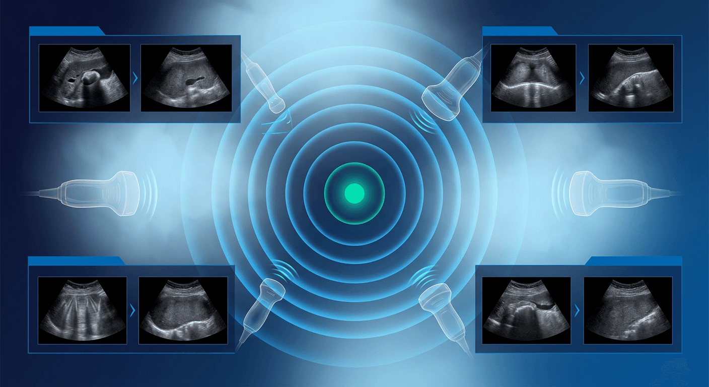
A complete abdominal ultrasound surveys the liver, gallbladder, bile ducts, pancreas, spleen, kidneys, and major abdominal vessels in one structured exam. For pain, abnormal laboratories, a suspected mass, or aneurysm, you need a reproducible protocol, measurements you can defend, and recognition of artifacts that mimic disease. This guide covers exam scope, organ-by-organ technique, commonly cited normal ranges, and pitfalls that change the report.
Abdominal Ultrasound Protocol: What the Exam Includes
A diagnostic complete abdominal ultrasound documents each organ in longitudinal and transverse planes so a later reader can confirm anatomy and calipers. Standard coverage includes the liver, gallbladder and extrahepatic bile duct, pancreas, spleen, both kidneys, the abdominal aorta to the bifurcation, and the inferior vena cava. Add the bladder, an ascites survey, and Doppler of the portal or hepatic veins when the indication requires them. Limited or bedside studies answer one question. A complete exam fits a broad differential, the same idea discussed in this overview of diagnostic versus point-of-care ultrasound. Typical indications include right upper quadrant or epigastric pain, abnormal liver enzymes or jaundice, organomegaly or unexplained ascites, flank pain or suspected hydronephrosis, and aortic aneurysm screening or surveillance.
Preparation, Positioning, and Transducer Choice
Fasting for about 6 to 8 hours reduces bowel gas and keeps the gallbladder distended. Small sips of water for medications are usually acceptable, and a full bladder is not required for a standard abdominal survey. Patients typically wear a gown or expose the abdomen from the xiphoid to the iliac crests. Scanning often takes about 20 to 30 minutes. Use a curved-array transducer in the low-frequency abdominal range, switching to a higher-frequency linear transducer for the gallbladder wall or superficial structures in thin patients. Start supine, then use oblique, lateral decubitus, and semi-erect positions when the gallbladder, pancreas, or left lobe is poorly seen.
Organ-by-Organ Scanning Technique
Liver
Sweep both lobes in sagittal and transverse planes. Document hepatic veins draining to the IVC, the portal vein, and parenchyma for echotexture, contour, and focal lesions. Compare hepatic echogenicity with the adjacent kidney. If you report length, measure the right lobe in a consistent midclavicular sagittal plane and state the method, because published upper limits vary with technique.
Gallbladder and bile ducts
Ultrasound of the gallbladder is most reliable after fasting. Image in at least two planes and change position, including left lateral decubitus and upright views, to show mobility of stones versus polyps or sludge. Measure wall thickness on the anterior wall, with the beam perpendicular, and avoid a contracted gallbladder. Follow the extrahepatic duct toward the pancreatic head. Look for shadowing calculi, sludge, wall thickening, pericholecystic fluid, a sonographic Murphy sign, and duct dilatation. Duodenal gas can hide the distal duct or mimic a stone, and edge shadowing at the neck should not be called a calculus. Case-based review of gallbladder on ultrasound is useful for less common wall and lumen findings.
Pancreas, spleen, and kidneys
Use the left hepatic lobe as a window to the pancreatic body, then sweep the head at the portal-splenic confluence and the tail toward the splenic hilum. A semi-erect position or a few sips of water can displace gas. Do not overcall a normal hypoechoic ventral anlage as a mass, and do not accept an unseen tail as a complete exam. Scan the spleen from a left intercostal window and measure maximum length in a consistent plane. For the kidneys, obtain true long-axis and transverse images, compare cortical echogenicity with liver and spleen, and document dilatation, calculi, cysts, and solid masses.
Abdominal aorta and IVC
Ultrasound for abdominal aorta evaluation follows the vessel from the celiac axis to the iliac bifurcation in sagittal and transverse planes. Measure anteroposterior diameter outer wall to outer wall, perpendicular to the aortic long axis, at the widest point. An aneurysm is commonly defined as 3.0 cm or greater. Oblique calipers overestimate diameter and cause follow-up disagreement. Assess the IVC for caliber, respiratory change, and thrombus when relevant.
Commonly Cited Normal Measurements
Cutoffs differ among sources. Report the value and plane, and prefer ranges over a single rigid number.
- Gallbladder wall: commonly 3 mm or less when fasting and not contracted
- Common bile duct: inner diameter often cited up to 6 mm, some sources use 7 mm; may increase with age and after cholecystectomy
- Pancreatic duct: often about 2 mm or less in the body, slightly larger in the head and with age
- Spleen length: commonly cited adult upper limits of about 12 to 13 cm
- Kidney length: commonly about 9 to 12 cm in adults
- Portal vein: often cited as less than 13 mm at the porta hepatis
- Abdominal aorta: 3.0 cm or greater is the usual aneurysm threshold; further management follows current vascular guidelines
Findings That Change Management
- Stones plus wall thickening, pericholecystic fluid, and a sonographic Murphy sign, supporting acute cholecystitis
- Dilated ducts, especially with a visible choledocholith or a pancreatic-head mass
- Aortic diameter at or above the aneurysm threshold, with location and maximum size stated
- Hydronephrosis with infection, rising creatinine, or suspected obstruction
- Unexplained free fluid, portal vein thrombosis, or a solid visceral mass needing further characterization
Ultrasound is first-line for cholelithiasis, many biliary questions, hydronephrosis, and aortic screening. Gas, habitus, and incomplete fasting limit the pancreas, distal duct, and retroperitoneum. When a required organ is not seen, state that it was incompletely evaluated.
Frequent Pitfalls in the Reading Room
- Calling wall thickening on a postprandial, contracted gallbladder
- Mistaking edge shadowing or duodenal gas for a shadowing stone
- Measuring the aorta off-axis or calling a tortuous vessel an aneurysm
- Foreshortening kidney or spleen length on an oblique image
- Overcalling reverberation in the gallbladder lumen as sludge
- Stopping the hepatic sweep at the gallbladder and missing a left-lobe lesion
Scan in the same sequence every time and store images with calipers on the structures you describe. IAME offers accredited ultrasound CME courses on abdominal and gallbladder imaging for structured case review.
Frequently Asked Questions
What can be detected on an abdominal ultrasound?
A complete study can show gallstones, cholecystitis, bile-duct dilatation, hydronephrosis, renal stones and masses, hepatic steatosis or cirrhotic texture, many focal liver and splenic lesions, ascites, and abdominal aortic aneurysm. It is less reliable for the pancreatic tail, bowel, and retroperitoneum when gas or body habitus limits the window. Cystic versus solid distinction is a strength, but CT or MRI is often needed for indeterminate masses.
How long does an abdominal ultrasound usually take?
A straightforward complete exam often takes about 20 to 30 minutes of scanning time. Limited windows, bowel gas, a large body habitus, or unexpected findings can extend the study. Distinguish scanning time from total department time when counseling patients.
Do you take your clothes off for an abdominal ultrasound?
Patients do not need to disrobe completely. A gown or lifting clothing enough to expose the abdomen from the xiphoid to the iliac crests is usually sufficient. Jewelry over the scanning field should be removed so it does not interfere with transducer contact.
Can you drink water before an abdominal ultrasound?
Most laboratories ask patients to fast for about 6 to 8 hours so the gallbladder remains distended and bowel gas is reduced. Small sips of water to take medications are commonly allowed. Confirm the local protocol, because some departments permit clear liquids and others prefer nothing by mouth except essential medications.