When a patient presents with a palpable lump, a focal mammographic finding, or dense tissue that limits mammography, ultrasound imaging breast evaluation is often the next practical step. High-frequency gray-scale and Doppler ultrasound can show whether a finding is a simple cyst, a probably benign solid mass, or a lesion that warrants biopsy. This scanner-ready guide covers indications, a systematic protocol, clock-face documentation, and the features that help separate benign from suspicious lesions.
When Breast Ultrasound Is the Right Next Study
Start with the clinical question. Targeted diagnostic ultrasound is appropriate for a new palpable mass, a mammographic mass or asymmetry that needs characterization, ultrasound correlation of an MRI lesion, a suspected intraductal mass with nipple discharge, implant assessment, and guidance for aspiration or core biopsy. Whole-breast screening ultrasound may be added for dense breasts as an adjunct, not a replacement for mammography.
- Palpable mass, thickening, or focal clinical concern
- Mammographic mass, asymmetry, or selected architectural distortion
- MRI-directed ultrasound for a potential ultrasound biopsy correlate
- Nipple discharge when an intraductal lesion is suspected
- Implant integrity and peri-implant fluid
- Supplemental screening in dense breasts
- Real-time guidance for cyst aspiration or percutaneous biopsy
Breast ultrasound versus mammography and MRI
Mammography remains the primary screening examination and the better test for calcifications and some architectural distortion. Ultrasound adds value for cystic versus solid characterization, dense tissue, and patients who are young, pregnant, or lactating. Breast ultrasound for cancer workups is strongest for masses and for planning a safe biopsy path. MRI has higher sensitivity for additional sites of disease and is used for high-risk screening, extent-of-disease evaluation, and problem solving when mammography and ultrasound are inconclusive. Do not assume every patient needs all three tests.
Ultrasound Imaging Breast Protocol: Positioning and Coverage
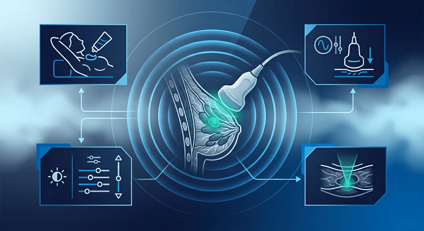
Place the patient supine with the ipsilateral arm abducted and the hand behind the head so the breast flattens on the chest wall. For outer-quadrant, axillary tail, and far-lateral lesions, roll the patient into a contralateral posterior oblique. On a targeted exam, scan the clinical or mammographic target first, then decide whether the remainder of the breast and axilla need coverage.
Use a high-frequency linear transducer and image every finding in two orthogonal planes, typically radial and antiradial or transverse and sagittal. Overlap survey rows on a whole-breast exam. Use lighter pressure in the retroareolar region so you do not empty small ducts or flatten a mobile mass. Survey the ipsilateral axilla when the indication is a new mass or known malignancy. For frequency, gain, and focal zone choices, see how to optimize breast ultrasound image quality.
Clock position, nipple distance, and depth
Annotate laterality, clock-face location, distance from the nipple, and depth on every key image. Each breast is its own clock with the nipple at the center. Twelve o'clock is superior and six o'clock is inferior. On the right breast, three o'clock is lateral and nine o'clock is medial. On the left breast, three o'clock is medial and nine o'clock is lateral. Measure centimeters from the nipple along that radius, then record depth from the skin. Mixing right and left conventions is a common source of wrong-site correlation.
A practical scanning checklist
- Confirm laterality and the mammographic, MRI, or clinical target before scanning.
- Palpate with the transducer so the palpable finding is the structure on the screen.
- Document the finding in two orthogonal planes and measure three diameters, including the longest axis.
- Apply color or power Doppler with a low scale to look for internal vessels.
- Record clock position, centimeters from the nipple, depth, and transducer orientation.
- State whether the finding is concordant with the mammogram or MRI location.
Gray-Scale Features: Normal Tissue Versus a Real Lesion
Normal breast ultrasound shows skin, subcutaneous fat, echogenic Cooper ligaments, mixed fibroglandular tissue, retromammary fat, and pectoralis muscle. Fat lobules are a frequent false mass: oval, parallel, isoechoic to fat, compressible, and without internal flow. If the structure blends into adjacent fat with light pressure, it is probably not a mass.
A simple cyst is anechoic and circumscribed, with an imperceptible wall, posterior enhancement, and no internal flow. Complicated cysts may contain homogeneous low-level echoes but should lack a mural nodule, thick wall, or vascularity. Once a finding is solid, describe shape, orientation, margin, echo pattern, and posterior features with consistent lexicon terms. Case-based introduction to breast imaging review can reinforce that lexicon.
Features that favor a benign solid mass
- Oval shape
- Parallel orientation, wider than tall
- Circumscribed margin
- Uniform mild hypoechogenicity or gentle hyperechogenicity
- Thin echogenic capsule
- Two or three gentle lobulations rather than microlobulation
- Mobility and compressibility
This pattern is typical of a fibroadenoma, but classic features still require concordance with the clinical exam and mammography. When the appearance is not classic, do not force a benign assessment.
Features that raise concern on breast cancer ultrasound imaging
- Irregular shape
- Nonparallel orientation, taller than wide
- Angular, microlobulated, or spiculated margins
- Thick echogenic halo
- Marked hypoechogenicity
- Posterior shadowing not explained by edge artifact
- Associated microcalcifications or intraductal extension
- Architectural distortion of surrounding tissue
No single feature proves malignancy. Some cancers can look oval and circumscribed. Combine gray-scale findings, Doppler, and multimodality correlation before choosing follow-up or biopsy.
Doppler, Measurements, and Correlation Pearls
Internal penetrating vessels in a solid mass are more concerning than absent or purely peripheral flow. Color inside a purported cyst means it is not a simple cyst. Absence of flow does not exclude cancer, especially in small or deep lesions. Measure three orthogonal diameters and use the same plane on follow-up. For MRI-directed ultrasound, remember the patient was prone for MRI and is supine for ultrasound, so clock position and depth can shift.
If you cannot find a reported mammographic or MRI mass, say so rather than substituting a nearby fat lobule.
Common Pitfalls at the Scanner
- Calling a fat lobule or ridge of glandular tissue a mass
- Missing far-lateral and axillary tail lesions because the breast was not rolled
- Overcompressing a mobile mass so it looks falsely parallel
- Pressing too firmly at the nipple-areolar complex and flattening a small retroareolar mass
- Omitting the axilla when the indication is a new mass or known cancer
- Annotating clock position without laterality
- Using ultrasound as a stand-alone substitute for screening mammography
From Imaging Finding to Next Step
Simple cysts that meet all criteria can be observed or aspirated for symptoms. Solid masses with classic probably benign features may be followed when concordant. Suspicious morphology needs tissue, usually ultrasound-guided core biopsy when the lesion is visible and approachable. Imaging-pathology concordance belongs in the same workflow. For targeting and sampling technique, see the course on ultrasound guided breast biopsy. If you also interpret dense-breast supplemental studies, a screening breast ultrasound curriculum can keep protocols consistent.
A disciplined protocol, complete annotation, and honest correlation with mammography and MRI will do more for interpretation than any single gray-scale sign. IAME offers accredited ultrasound CME courses on breast imaging for clinicians who want additional case review on this topic.
Frequently Asked Questions
Do ultrasounds show breast cancer?
Yes. Many invasive cancers appear as irregular, nonparallel, or spiculated solid masses, and ultrasound is a standard test for characterizing a lump or a mammographic finding. Ultrasound is less sensitive for some ductal carcinoma in situ that presents only as calcifications, which mammography detects more reliably. A normal targeted ultrasound does not exclude malignancy if the mammogram or clinical exam remains unexplained.
Are a mammogram and a breast scan the same?
No. Mammography uses ionizing radiation and compression. Breast ultrasound uses sound waves without ionizing radiation and is usually targeted to a symptom or an imaging finding. The tests are complementary, not interchangeable. Breast scan is an informal phrase that can mean ultrasound, MRI, or another study, so it is clearer to name the modality.
How should clock position be documented on breast ultrasound?
Treat each breast as its own clock with the nipple at the center and twelve o'clock toward the head. State laterality first, then clock position, distance from the nipple in centimeters, and depth from the skin. Include transducer orientation on the annotation so another examiner can reproduce the finding.
Is breast ultrasound useful for dense breasts?
Yes, as a supplemental test. Dense tissue can mask masses on mammography, and ultrasound can show masses that mammography does not. Supplemental ultrasound also increases biopsies of benign lesions. It does not replace mammography, which remains the better test for calcifications and some architectural distortion.
Can a breast cancer ultrasound scan replace mammography?
No. A breast cancer ultrasound scan is excellent for mass characterization, young or pregnant patients, and biopsy guidance, but it is not a stand-alone screening test for most patients. Mammography remains the primary screening examination. Use ultrasound to answer a specific diagnostic question or as an adjunct in selected settings such as dense breasts.