Discover insights, updates, and educational content from our medical education experts. Stay informed with the latest trends and developments in healthcare education.
Loading…
Connect with Us!
Feel free to reach out to us for further information!
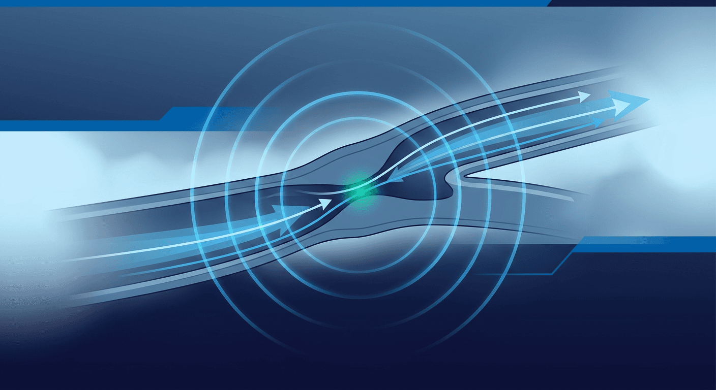
Top 5 Vascular Ultrasound Techniques for Better Results
By Jason Cox, MD
Top 5 Vascular Ultrasound Techniques for Better Results
Key Techniques:
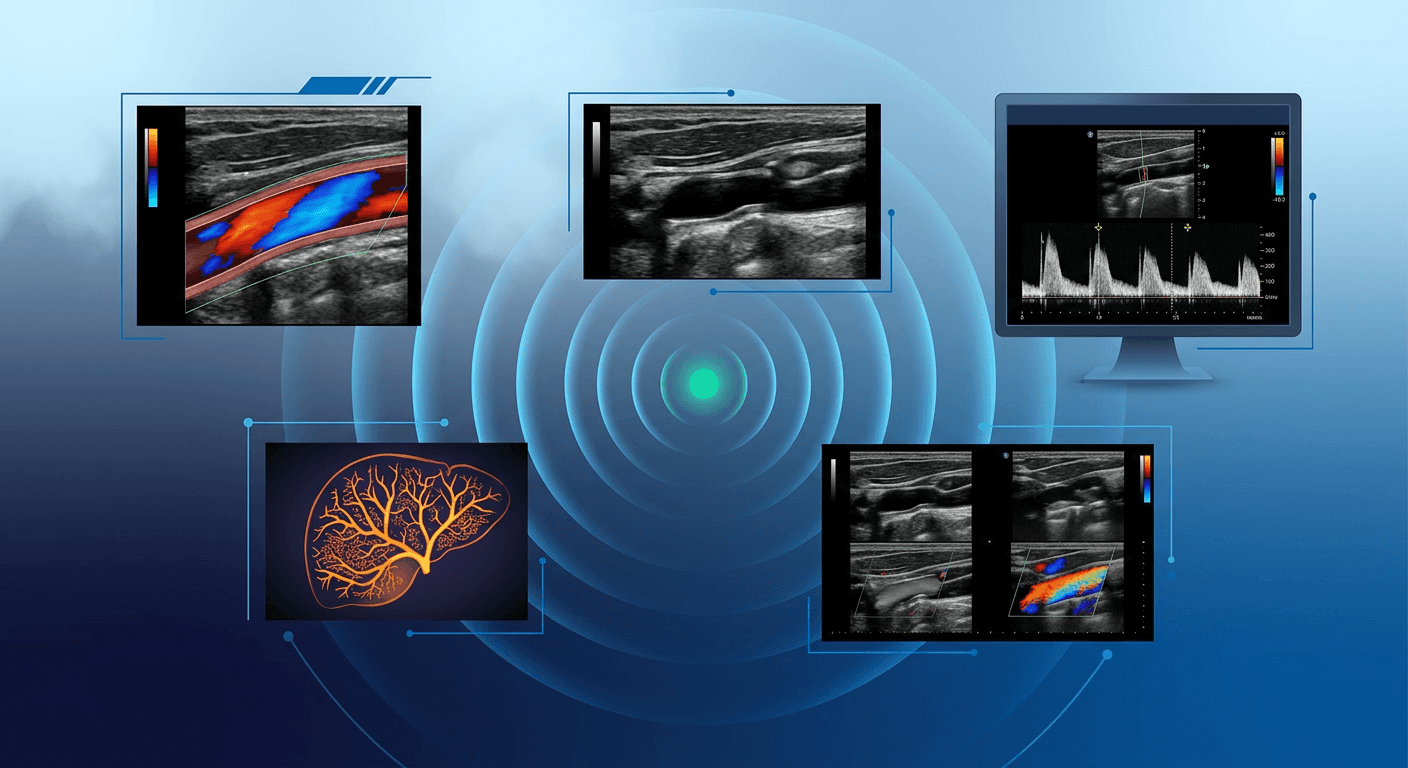
Doppler Imaging: Advanced blood flow analysis with methods like Color, Power, Spectral, and Duplex Doppler.
Linear Array Scanning: High-resolution imaging for detailed vessel assessments.
Contrast-Enhanced Ultrasound (CEUS): Microbubble agents for enhanced blood flow visualization, detecting vessels as small as 40 μm.
3D Vessel Imaging: Spatial views of blood vessels for better analysis of complex anatomy.
Image-Guided Procedures: Real-time visualization for precise interventions.
Quick Comparison:
Technique
Best For
Advantage
Drawback
Doppler Imaging
Blood flow analysis
Real-time insights
Requires angle adjustments
Linear Array Scanning
Vessel structure imaging
High resolution for shallow vessels
Limited depth penetration
CEUS
Low-flow detection
Detects microvascular flow
Requires contrast agents
3D Vessel Imaging
Complex anatomy analysis
Submillimeter accuracy
Specialized equipment needed
Image-Guided Procedures
Interventional procedures
Precision and reduced errors
Requires advanced training
These techniques improve diagnostic accuracy, enhance imaging precision, and support better clinical outcomes. Dive into the article for detailed insights on how to apply them effectively.
Vascular ultrasound basics
1. Doppler Imaging Techniques
Modern Doppler imaging offers advanced methods for analyzing blood flow, addressing challenges like detecting low-flow states mentioned earlier.
Color Doppler uses red and blue mapping to show blood flow direction, making it useful for spotting issues like stenosis or thrombosis[5][7]. It helps overcome the limitations of B-mode imaging, especially in distinguishing blockages from low-flow conditions.
Power Doppler improves sensitivity to slow-moving blood, making it ideal for examining microvascular structures or detecting low-flow states that traditional methods might miss. For example, it can identify residual flow in partially blocked vessels that other techniques might fail to detect[5].
Doppler Method
Main Use
Advantage
Drawback
Color Doppler
Shows blood flow direction
Real-time visualization
Less sensitive to slow flow
Power Doppler
Examines microvascular flow
Detects slow-moving blood
Doesn't show flow direction
Spectral Doppler
Measures blood velocity
Offers precise flow data
Limited to single points
Duplex Doppler
Combines imaging and flow data
Provides detailed analysis
Needs a skilled operator
Spectral Doppler focuses on measuring blood flow velocity. Pulsed wave (PW) Doppler targets specific locations, while continuous wave (CW) Doppler captures higher velocities, making it versatile for various clinical needs.
Duplex Doppler combines B-mode imaging with Doppler analysis, offering both anatomical and blood flow insights in a single technique[2]. This integration provides a more detailed and comprehensive view for diagnostics.
For the best results, operators should keep the Doppler angle at 60 degrees or lower relative to blood flow. Adjusting gain and pulse repetition frequency helps minimize artifacts like aliasing and blooming, which can affect image clarity.
Newer tools, such as Vector Flow Imaging (VFI), are now enhancing these methods by removing angle dependency[8]. These innovations improve diagnostic accuracy and align with SVU guidelines for better clinical practices.
2. Linear Array Scanning Methods
Linear array transducers play a key role in vascular ultrasound, featuring 256-512 elements arranged precisely. These devices provide lateral resolution of under 0.5 mm, allowing for detailed imaging of vessel structures and accurate vessel diameter measurements[6]. This precision is particularly useful for addressing the challenges of intima-media thickness measurements discussed earlier.
High-Resolution Imaging Modern linear arrays offer a rectangular field of view, making them ideal for vascular assessments. They maintain uniform beam density across the scanning field, aligning with SVU guidelines for standardized vascular evaluations[3].
Frequency Applications:
Frequency
Best For
Advantage
7-15 MHz
Superficial vessels (<4 cm)
High resolution for carotid arteries
3-7 MHz
Deeper structures
Improved penetration for larger patients
2-8 MHz
General vascular assessments
Broad usability across different needs
Advanced Technologies Beyond Doppler flow analysis, modern linear arrays now include spatial compounding, which combines multiple beam angles to minimize artifacts and improve tissue differentiation[3]. Tissue harmonic imaging further enhances contrast resolution, especially helpful for patients with dense or thick tissue, such as those with obesity[1].
Practical Tips Adjust the gain carefully to make vessel walls clear without oversaturating the image[1].
For example, the BK 8L2 transducer showcases these advancements with:
A 45 x 14 mm contact surface
A sterilizable design
Want to keep learning? Our CME memberships make it easy with unlimited access to accredited courses year-round.
3. Contrast-Enhanced Ultrasound (CEUS)
Contrast-Enhanced Ultrasound (CEUS) has transformed vascular imaging by using microbubble contrast agents to improve blood flow visualization. These microbubbles, which are smaller than red blood cells, significantly enhance image clarity and diagnostic precision compared to standard ultrasound techniques[4]. This method effectively addresses the challenges of detecting low blood flow, a limitation of conventional Doppler imaging.
Key Clinical Applications
CEUS tackles two major challenges: distinguishing blocked vessels and achieving submillimeter precision. It is particularly effective in the following areas:
CEUS provides real-time insights into blood flow and perfusion, making it invaluable for complex vascular cases. It offers better visualization of vessel walls and allows live monitoring of blood flow dynamics[10]. These features address many of the limitations found in standard B-mode imaging.
Practical Implementation
To use CEUS effectively, specialized ultrasound systems with contrast-specific imaging modes are required. For best results, clinicians should:
Select the appropriate contrast agent for the specific exam.
Follow established protocol templates.
Verify renal status, as these agents are safe for kidneys.
This approach builds on traditional Doppler methods, eliminating issues like angle dependency.
Clinical Benefits
CEUS stands out as a superior option for monitoring vascular conditions, such as after endovascular aneurysm repair, where consistent follow-ups are crucial.
"CEUS can detect blood flow in vessels as small as 40 μm in diameter, far beyond the capabilities of conventional Doppler ultrasound[4]."
Recent Developments
The technology continues to advance with the addition of 3D imaging and artificial intelligence. These tools improve diagnostic precision and streamline workflows[9]. For instance, Q-CEUS provides measurable data on tissue perfusion, aiding clinical decision-making[4].
Safety Profile
With a severe reaction rate of less than 0.01%[2], CEUS offers a safer alternative for repeat imaging compared to contrast-enhanced CT scans.
If you'd like to keep learning, stay up to date, and improve patient outcomes, learn more here.
4. 3D Vessel Imaging
Expanding on the insights provided by CEUS, 3D vessel imaging introduces a spatial perspective to vascular analysis. This is especially useful for examining low-flow vascular networks and borderline stenosis cases. With this method, clinicians can generate three-dimensional views of blood vessels, allowing for analysis from various angles.
Advanced Reconstruction Methods
Modern 3D vessel imaging uses a variety of sophisticated techniques for data collection and reconstruction:
Method
Key Features
Clinical Applications
Matrix Array Transducers
Captures real-time volumetric data
Evaluating complex vessel anatomy
Mechanical 3D Probes
Performs automated volume sweeps
Planning surgeries for intricate bypass grafts
Freehand Scanning
Tracks 2D image sequences
Mapping large vessels
Fusion Imaging
Combines ultrasound with CT or MRI data
Pre-operative planning
Clinical Accuracy and Performance
The use of 3D vessel imaging has significantly enhanced diagnostic precision, achieving submillimeter accuracy for plaque burden assessments. Studies show that 3D ultrasound detects carotid artery stenosis with a 93% accuracy rate, compared to 77% with traditional 2D ultrasound [3]. This directly addresses the challenges of intima-media thickness measurements discussed earlier. Additionally, volumetric plaque measurements via 3D imaging offer a reproducibility rate of ±3.1%, far exceeding the ±11.8% rate seen with 2D methods [11].
Practical Applications
A 2022 study by Dr. Michael Lee at the University of California, San Francisco, highlighted the real-world impact of 3D vascular ultrasound. In a group of 150 patients with carotid artery disease, the technology identified 12% more significant stenoses than conventional methods. This led to treatment changes for 8% of the patients [12].
Technical Considerations
To achieve the best results with 3D vessel imaging, clinicians should:
Capture images from multiple angles while maintaining stable positioning.
Fine-tune B-mode settings before starting the scan.
Use breath-hold techniques for imaging abdominal vessels.
"3D ultrasound can create virtual 'fly-through' views of blood vessels, providing unmatched visualization of vascular structures[2]."
Integration with Other Techniques
3D imaging pairs effectively with Doppler for volumetric flow mapping and AI tools for plaque analysis. Recent advancements include high-frequency matrix array transducers and enhanced image processing algorithms, pushing diagnostic capabilities even further [8].
5. Image-Guided Procedures
Using 3D spatial visualization, real-time image guidance brings accuracy to vascular ultrasound applications. Ultrasound-guided procedures bridge diagnostics and treatment, aligning with SVU guidelines to promote consistent care.
This approach leverages the high resolution of modern linear arrays (discussed in section 2) for therapeutic purposes. Tools like echogenic needles and electromagnetic tracking systems have helped cut procedure times by 30% [7].
Clinical Applications and Outcomes
Ultrasound guidance has been shown to lower central line infection rates by 57% and boost first-attempt IV success rates to 80-90% [5].
Quality Assurance Programs
To maintain high standards, quality assurance measures include:
Competency Evaluations: Skills are tested through simulation-based assessments.
Outcome Monitoring: Digital systems track procedure success rates.
Technical Considerations
Achieving the best results requires sterile techniques, selecting the right frequency for depth, and using Doppler for verification [2][5].
Pre-procedure Planning
Successful interventions depend on several key steps:
Vessel Assessment: High-frequency linear transducers provide detailed imaging of target vessels.
Real-time Visualization: Keeping the needle tip visible during advancement reduces accidental punctures by 71% [5].
Follow-up Monitoring: Structured post-procedure checks help detect complications early [7].
Method Comparison
Different vascular ultrasound techniques come with their own pros and cons, helping clinicians address diagnostic challenges like low-flow states and precision issues. These differences play a critical role in shaping clinical decisions for the vascular issues discussed earlier.
Resolution and Depth Capabilities
Choosing the right technique often depends on the trade-off between depth and resolution. For instance, linear arrays work well for shallow imaging (<6cm) with high detail, while Doppler ultrasound has limited depth penetration. 3D vessel imaging offers a broader spatial view but at the cost of some resolution, whereas contrast-enhanced ultrasound (CEUS) improves visualization at all depths [4].
Technical Implementation
Applying SVU guidelines requires balancing the technical features of each method with practical considerations like cost and ease of use:
Technique
Cost Factors
Learning Curve
Doppler
Low equipment
Moderate
Linear Array
Standard
Moderate
CEUS
High (contrast)
Steep
3D Imaging
Specialized
High
Image-Guided
Advanced systems
Extensive
Future Integration
Emerging technologies, especially AI, are being integrated to refine vessel analysis [12]. Combining multiple techniques is becoming more common, particularly in complex cases, to improve diagnostic precision and treatment planning [3].
Clinical Impact
Most methods are non-invasive and cause little discomfort. However, CEUS involves administering contrast agents, and image-guided procedures require advanced technical skills [4]. This comparison helps clinicians use the techniques discussed in Sections 1-5 more effectively in everyday practice.
"3D ultrasound can create virtual 'fly-through' views of blood vessels, providing unmatched visualization of vascular structures [2]."
Conclusion
The five techniques discussed - ranging from Doppler advancements to 3D imaging - tackle some of the biggest challenges in modern vascular imaging seen in clinical practice. Studies show these methods improve diagnostic accuracy by 20-30% compared to traditional ultrasound, especially in low-flow states and borderline stenosis cases[2].
The Society for Vascular Ultrasound (SVU) has updated its certification protocols to include these methods, ensuring consistent use across facilities[3]. Clinical data highlights that centers adopting these techniques report a 23% drop in diagnostic errors for stenosis grading[11].
To apply these methods effectively, sonographers need robust quality assurance systems and ongoing skill development. When paired with structured documentation and outcome tracking, these techniques allow professionals to:
Measure vessels with submillimeter precision
Identify low-flow states that older methods often miss
Offer more accurate guidance during interventional procedures
By consistently applying these evidence-based methods, sonographers can deliver more accurate assessments and better-informed treatment decisions. Combining tools like Doppler imaging, linear array scanning, CEUS, 3D vessel imaging, and image guidance creates a well-rounded approach to vascular diagnostics, directly addressing the challenges outlined earlier.
"3D ultrasound can create virtual 'fly-through' views of blood vessels, providing unmatched visualization of vascular structures[2]."
FAQs
What are the advances in vascular ultrasound?
Expanding on the core techniques mentioned earlier, some notable advances include:
Volumetric 3D Imaging for detailed plaque analysis (see Section 4)
AI-powered Doppler methods (see Section 1)
Enhanced contrast protocols (see Section 3)
These developments significantly improve 3D vessel imaging and Doppler functionality, building on the foundational techniques previously discussed.
What affects image quality in ultrasound?
Several factors play a role in determining the quality of ultrasound images. Here's a quick overview:
Factor
Impact on Image Quality
Tissue Composition
Influences how well beams penetrate
Depth
Can lower resolution
Transducer Choice
Impacts imaging precision
Patient Anatomy
Affects accessibility
For example, selecting the right transducer (see Section 2) helps address depth-related issues, while advanced imaging techniques can mitigate anatomical challenges.
To improve image quality, consider these steps:
Ensure your system is properly calibrated
Adjust scanning angles to match vessel anatomy
Use AI-based tools for noise reduction and enhanced clarity
AI-driven noise reduction is particularly useful, offering clearer images to support accurate vascular diagnoses [13].
Courses on IAME integrate ultrasound physics and artifacts into the learning. Learn more about artifacts when you get an all-access membership to IAME today.
If you'd like unlimited access to CME for years to come, take a look at our memberships here. As a thank-you, use code POST at checkout to save 12% on any membership.
If you're a physician, we recommend checking out this CME program.
 If you need any assistance with your CME, we're here to help. Just email us at