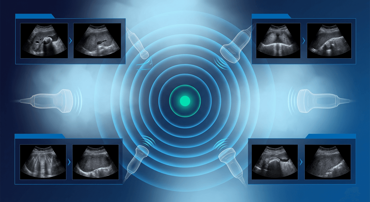
Ultrasound artifacts can distort medical images, leading to misdiagnoses if not properly addressed. This guide explains common artifacts like acoustic shadowing, reverberation, tissue-related issues, and motion artifacts, along with practical solutions to improve image quality and diagnostic accuracy.
Key Points:
Acoustic Shadowing: Caused by dense structures blocking sound waves. Adjust transducer angle, pressure, or use alternative imaging techniques.
Reverberation Effects: Occur due to repeated sound wave reflections. Modify insonation angle, lower near-field gain, and use advanced imaging methods.
Tissue-Related Artifacts: Result from density variations. Optimize machine settings like frequency, gain, and focus zone.
Motion and Blood Flow Artifacts: Caused by patient or transducer movement. Encourage breath-holding, adjust Doppler settings, and reduce color gain.
Quick Tips:
Use higher frequencies for shallow imaging, lower for deeper areas.
Enable harmonic imaging to reduce noise.
Adjust PRF and insonation angle for accurate blood flow measurements.
Understanding and correcting artifacts ensures better diagnostic outcomes and patient care.
If you'd like to keep learning, stay up to date, and improve patient outcomes, learn more here.
Acoustic Shadowing
How Shadowing Happens
Acoustic shadowing is an ultrasound artifact that appears when sound waves hit dense or highly reflective structures. These structures - like bone, calcifications, or air interfaces - block the sound waves, creating a dark "shadow" on the image beneath them.
When ultrasound waves encounter objects with high acoustic impedance, such as gallstones or bone, a significant amount of the sound energy is either reflected back or absorbed. This prevents the waves from passing through, leading to the shadow effect. Recognizing this phenomenon is key to improving image clarity and avoiding diagnostic errors.
Tips to Prevent Shadowing
To minimize shadowing, consider these practical steps:
Transducer Positioning: Keep the transducer perpendicular to the area being scanned for clearer images.
Pressure Control: Apply gentle, consistent pressure with the transducer to avoid tissue distortion and maintain proper blood flow.
Focal Depth Setting: Adjust the focal depth carefully. Shadows tend to be more noticeable when the focal depth is close to the obstructing object.
Techniques to Minimize Shadowing
If shadowing becomes a challenge, try these methods:
Interestingly, while shadowing often complicates imaging, it can also be diagnostically helpful. For example, an acoustic shadow behind the gallbladder can indicate the presence of gallstones, even if the stones themselves are too small to see directly [2].
Want to keep learning? Our CME memberships make it easy with unlimited access to accredited courses year-round.
Ultrasound Artifacts, Part 1 (Shadowing, enhancement and ...
Reverberation Effects
Managing reverberation artifacts is crucial for achieving clear ultrasound images and ensuring accurate diagnoses.
Types of Reverberation
Reverberation artifacts occur when ultrasound waves repeatedly bounce between reflective surfaces, producing multiple echo lines that can obscure anatomical details [3].
Comet tail artifacts: These appear as a short series of echoes originating from a highly reflective structure [4].
Ring-down artifacts: These resemble comet tail artifacts but are caused by a different mechanism [4].
Reducing Reverberation
To minimize the impact of these artifacts, consider the following techniques:
Transducer adjustments: Change the insonation angle to reduce parallel reflections [4].
Optimizing machine settings: Lower near-field TGC (Time Gain Compensation) to help reduce comet tail artifacts [5].
Advanced imaging methods: Techniques like M-mode echocardiography and multiplane transesophageal echocardiography (TEE) can help distinguish artifact types and confirm the accuracy of images [5].
Tissue Effects on Images
Tissue density plays a key role in ultrasound image quality and how well sound waves travel through the body. To improve imaging and reduce artifacts like shadowing and reverberation, understanding how tissues interact with ultrasound waves is crucial. Let’s explore how tissue variations impact imaging and how machine settings can be adjusted to address these challenges.
Tissue Density Effects
Ultrasound waves behave differently when they pass through tissues with varying densities [1]. This interaction can create artifacts that influence diagnostic accuracy.
Dense tissues, such as bone or calcifications, reflect most of the ultrasound waves, limiting penetration.
Fluid-filled areas allow waves to pass through more easily, improving image clarity.
These tissue differences affect:
Signal strength and how deep the waves can penetrate
Image clarity, including resolution
Artifact patterns, which may obscure details
Overall diagnostic precision
Spotting Image Artifacts
Artifacts caused by tissue variations can be identified with careful observation [6]. Watch for:
"During their training, sonographers are taught, and encouraged, to use the minimum amount of acoustic power to obtain a quality image. The common term is ALARA (As Low As Reasonably Achievable)." - Innovatus Imaging [7]
Machine Setting Adjustments
Adjusting ultrasound machine settings can help minimize the impact of tissue-related artifacts and improve image quality. Here’s a quick guide:
Parameter | Adjustment | Effect |
|---|
Frequency | Use the highest possible for the region of interest (ROI) | Improves resolution but limits depth |
Harmonics | Enable Tissue Harmonic Imaging (THI) | Reduces noise in complex cases |
Dynamic Range | Lower for echo, higher for radiology | Enhances contrast resolution |
Compound Imaging | Enable CrossBeam/SonoCT | Better visualization of shadowed areas |
Speckle Reduction | Enable SRI/XRES/uSca | Reduces noise in fluid-dense structures |
For best results when scanning through tissues with varying densities:
Frequency Selection: Use higher frequencies for shallow targets and lower frequencies for deeper areas.
Focus Zone: Set the focal zone at or just beneath the area of interest for sharper imaging.
Gain Adjustments: Modify gain and time gain compensation (TGC) to balance tissue attenuation and improve visibility.
Motion and Blood Flow Artifacts
Motion and blood flow artifacts can reduce the accuracy of diagnostic imaging. Recognizing their causes and adjusting techniques can help improve results. Similar challenges arise in Doppler exams, impacting flow measurements.
Causes of Motion Artifacts
Motion artifacts can show up as blurring, streaking, or shading in ultrasound images, sometimes mimicking pathological conditions and obscuring important details [8]. These artifacts often stem from:
Patient Movement: Voluntary actions like breathing or shifting during the scan.
Physiological Motion: Involuntary movements, such as those caused by heartbeat, breathing, or blood vessel pulsation.
Transducer Movement: Unsteady handling of the ultrasound transducer.
Blood Flow Measurement Challenges
Doppler settings can also introduce issues with blood flow measurements. One common problem is aliasing, which happens when the pulse repetition frequency (PRF) is too low compared to the Doppler shift [9].
Here are some common challenges in blood flow measurement:
Issue | Cause | Result |
|---|
Aliasing | Low PRF | Color inversion and peak cutoff |
Blooming | Excessive color gain | Overestimated vessel diameter |
Spectral Broadening | Improper gate sizing | Unclear velocity measurements |
Addressing Motion and Flow Issues
To improve imaging quality and accuracy, try these adjustments:
Reducing Motion Artifacts: Encourage patients to hold their breath during critical scans. Use faster scanning techniques or cardiac gating when necessary [8].
Optimizing Doppler Settings: For better blood flow measurements, lower the insonation angle to under 60°, set the sample volume to about two-thirds of the vessel diameter, and increase the PRF when aliasing occurs [11].
Adjusting Color Doppler: Lower the color gain, tweak wall filters, and correct the baseline to enhance flow visualization [10].
For vascular access sampling, where high velocities can cause aliasing, increasing the PRF or using Continuous Wave Doppler can improve accuracy [9][11].
Summary
Managing ultrasound artifacts effectively is crucial for accurate diagnoses. Below, you'll find an overview of common imaging artifacts, their causes, and practical solutions.
Quick Reference Guide
Artifact Type | Common Causes | Prevention/Solution |
|---|
Acoustic Shadowing | Dense structures, calcifications | Adjust the transducer angle, reposition as needed, or use a deep transgastric long-axis view for cardiac imaging |
Reverberation | Multiple echo paths, strong reflectors | Lower near-field gain, change the beam angle, and verify with M-mode |
Motion | Patient movement, physiological activity | Use faster scanning techniques, apply cardiac gating, or ask the patient to hold their breath |
Blood Flow | Incorrect PRF settings, excessive gain | Adjust velocity scale to 15–35 cm/s, reduce color gain, and fine-tune the gate size |
Side-Lobe | Off-axis beam artifacts | Switch to harmonic imaging and adjust focal zones |
CME Resources at IAME

IAME provides ACCME-accredited courses designed to help healthcare professionals master ultrasound artifact recognition and management. The curriculum includes:
Advanced techniques for reducing artifacts
Optimizing machine settings for various clinical scenarios
Hands-on workshops for practical learning
For reference, here’s a frequency guide based on imaging depth:
Depth (cm) | Optimal Frequency (MHz) | Common Applications |
|---|
<2.0 | 12–15 | Superficial structures |
2.0–4.0 | 10–12 | Most peripheral blocks |
>4.0 | 5–10 | Deep tissue imaging |
Courses on IAME integrate ultrasound physics and artifacts into the learning. Learn more about artifacts when you get an all-access membership to IAME today.
If you'd like unlimited access to CME for years to come, take a look at our memberships here. As a thank-you, use code POST at checkout to save 12% on any membership.
If you're a physician, we recommend checking out this CME program.
If you're a sonographer, take a look at this one; we think you'll love it.
 If you need any assistance with your CME, we're here to help. Just email us at support@iame.com