Ultrasound Assessment and Mapping of the Superficial Venous System
0% Complete
Course Overview
This course provides a comprehensive guide to the ultrasound techniques used for assessing the superficial venous system, crucial for procedures such as bypass grafting and hemodialysis access fistula creation. Learn how to evaluate veins like the great and small saphenous veins, as well as the cephalic and basilic veins, by mapping their course, diameter, and patency. Key topics include the identification of normal venous structures, common anatomical variations, and potential issues such as thrombus, varicosities, and calcifications. This course is ideal for clinicians performing pre-operative venous assessments for surgical procedures.
Objectives
After completing this activity, the participant will:
Describe the normal anatomical features of the great saphenous vein, small saphenous vein, cephalic vein and basilic vein
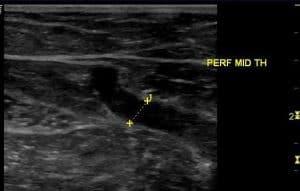
Identify pathology observed within the superficial veins
Describe the basic techniques of venous mapping
Identify limitations encountered during vein mapping
Describe diagnostic ultrasound criteria utilized in venous mapping
Target Audience
Physicians, sonographers, and others who perform and/or interpret ultrasound.
Faculty & Disclosure
Faculty
Ann Marie Kupinski, PhD RVT RDMS
Clinical Associate
Professor of Radiology, Albany Medical College, Albany NY
Technical Director,
North Country Vascular Diagnostics, Inc, Altamont NY
Disclosure
In compliance with the Essentials and Standards of the ACCME, all individuals in control of the content of this CME tutorial are required to disclose any significant financial or other relationships they may have with commercial interests.</p> <p>Ann Marie Kupinski, PhD RVT RDMS discloses no such relationships exist. No one at IAME who had control over the planning or content of this activity<br /> has relationships with commercial interests.
Credits
* AMA PRA Category 1™ credits are used by physicians and other groups like PAs and certain nurses. Category 1 credits are accepted by the ARDMS, CCI, ACCME, and Sonography Canada.
Course Details
Accreditation
The Institute for Advanced Medical Education is accredited by the Accreditation Council for Continuing Medical Education (ACCME) to provide continuing medical education for physicians.
The Institute for Advanced Medical Education designates this enduring material for a maximum of 2 AMA PRA Category 1 Credit™.
Physicians should only claim credit commensurate with the extent of their participation in the activity. Sonographers: These credits are accepted by the American Registry for Diagnostic Medical Sonography (ARDMS), Sonography Canada, Cardiovascular Credentialing International (CCI), and most other organizations.

