The Sonographic Detection Of Uterine Anomalies
0% Complete
Course Overview
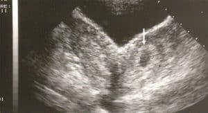
Explore the critical role of sonography in diagnosing uterine anomalies, a key factor in fertility and pregnancy outcomes. This course covers the identification and classification of uterine malformations such as unicornuate, didelphys, bicornuate, septate, and arcuate uteri, including how to distinguish between them using 2D and 3D ultrasound imaging. Learn the impact of uterine anomalies on pregnancy, including risks like miscarriage, preterm delivery, and cervical incompetence. Through detailed protocols, you'll gain expertise in visualizing and diagnosing abnormalities using 3D ultrasound, including the evaluation of uterine septa, bicornuate uteri, and more. This course is designed for clinicians looking to improve detection rates and enhance their understanding of the implications for reproductive health. Gain knowledge of the AFS classification system, diagnostic criteria, and advanced techniques for accurate assessment of uterine malformations.
Objectives
After completing this activity, the participant will:
Describe the classification of muellerian anomalies to major uterine anatomic types as defined by the American Fertility Society.
List the prevalence, complications and methods to detect uterine anomalies.
Apply transvaginal sonography and/or 3D ultrasound to distinguish specific uterine malformations.
Target Audience
Physicians, sonographers, and others who perform and/or interpret obstetrical ultrasound.
Faculty & Disclosure
Faculty
Lyndon M. Hill, MD
Professor Obstetrics and Gynecology
Medical Director Ultrasound
Magee Women's Hospital
Pittsburgh, PA
Disclosure
In compliance with the Essentials and Standards of the ACCME, the author of this CME tutorial is required to disclose any significant financial or other relationships they may have with commercial interests. Dr. Lyndon Hill discloses no such relationships exist. No one at IAME who had control over the planning or content of this activity has relationships with commercial interests.
In compliance with the Essentials and Standards of the ACCME, the author of this CME tutorial is required to disclose any significant financial or other relationships they may have with commercial interests.
IAME has assessed conflict of interest with its faculty, authors, editors, and any individuals who were in a position to control the content of this CME activity. Any identified relevant conflicts of interest have been mitigated. IAME's planners, content reviewers, and editorial staff disclose no relationships with ineligible entities.
Credits
* AMA PRA Category 1™ credits are used by physicians and other groups like PAs and certain nurses. Category 1 credits are accepted by the ARDMS, CCI, ACCME, and Sonography Canada.
Course Details
Accreditation
The Institute for Advanced Medical Education is accredited by the Accreditation Council for Continuing Medical Education (ACCME) to provide continuing medical education for physicians.
The Institute for Advanced Medical Education designates this enduring material for a maximum of 1 AMA PRA Category 1 Credit™. Physicians should only claim credit commensurate with the extent of their participation in the activity.
Sonographers: These credits are accepted by the American Registry for Diagnostic Medical Sonography (ARDMS), Sonography Canada, Cardiovascular Credentialing International (CCI), and most other organizations.

