Sonographic Evaluation of the Normal and Abnormal Placenta
0% Complete
Course Overview
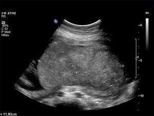
This course focuses on the sonographic evaluation of the normal and abnormal placenta, offering insights into placental development, morphology, and pathologies. Placental conditions such as previa, accreta, abruption, and chorioangiomas are examined, with emphasis on the role of ultrasound in detecting these abnormalities. The course includes specific techniques for identifying various placental conditions, including the use of color Doppler to assess blood flow in lesions like chorioangiomas and to detect complications such as fetal hydrops or polyhydramnios. It also covers the impact of placental abnormalities on fetal development, especially in cases like intrauterine growth restriction (IUGR). The curriculum also highlights the relationship between placental dysfunction and common complications in pregnancy, such as preeclampsia and elevated maternal serum alpha-fetoprotein levels.
Objectives
After completing this activity, the participant will:
Describe the development of the normal placenta and its' sonographic appearance.
Identify the changes that occur in the placenta with intrauterine growth restriction.
Apply diagnostic ultrasound to identify placenta previa, abruption and other abnormalities that are the potential source of fetal disease and / or compromise.
Target Audience
Physicians, sonographers, and others who perform and/or interpret obstetrical ultrasound.
Faculty & Disclosure
Faculty
Lyndon M. Hill, MD
Professor Obstetrics and Gynecolog
Medical Director Ultrasound
Magee Women's Hospital
Pittsburgh, PA
Disclosure
In compliance with the Essentials and Standards of the ACCME, the author of this CME tutorial is required to disclose any significant financial or other relationships they may have with commercial interests. Dr. Lyndon M. Hill has indicated that no such relationships exist. No one at IAME who had control over the planning or content of this activity has relationships with commercial interests.
Credits
* AMA PRA Category 1™ credits are used by physicians and other groups like PAs and certain nurses. Category 1 credits are accepted by the ARDMS, CCI, ACCME, and Sonography Canada.
Course Details
Accreditation
The Institute for Advanced Medical Education is accredited by the Accreditation Council for Continuing Medical Education (ACCME) to provide continuing medical education for physicians.
The Institute for Advanced Medical Education designates this enduring material for a maximum of 1 AMA PRA Category 1 Credit™.
Physicians should only claim credit commensurate with the extent of their participation in the activity. Sonographers: These credits are accepted by the American Registry for Diagnostic Medical Sonography (ARDMS), Sonography Canada, Cardiovascular Credentialing International (CCI), and most other organizations.

