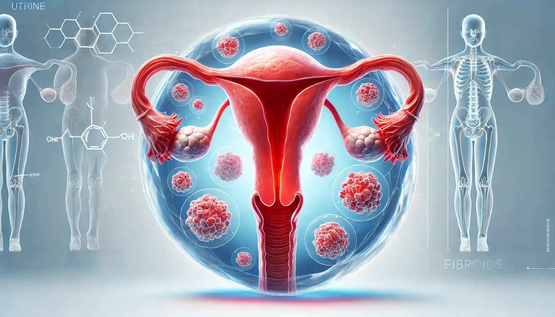
Uterine Fibroids
0% Complete
Course Overview
Course Content
Credits
* AMA PRA Category 1™ credits are used by physicians and other groups like PAs and certain nurses. Category 1 credits are accepted by the ACCME, ARDMS, CCI, and Sonography Canada.
Course Details
- Duration
- 1.00 hour
- Released
- Mar 1, 2023
- Last Review
- Dec 9, 2024
- Expires
- Dec 8, 2027
Objectives
After completing this activity, the participant will:
Perform a complete sonographic examination for evaluation
Understand the various sonographic characteristics
Understand the various anatomical locations of uterine fibroids
Target Audience
Physicians, sonographers, and others who perform and/or interpret ultrasound.
Faculty & Disclosure
Faculty
Lauren Pellegrini, MHA, RDMS, RVT, RDCS
Diagnostic Medical Sonography Program Director, St. Louis Community College-Forest Park
Sonographer and Clinical Sonography Instructor
St. Louis, MO
Jason Cox, MD
Program Director, Medical Director, and Attending Radiologist
Institute for Advanced Medical Education
St. Louis, MO
Christopher J. Allen RVT, RDMS, (AB) (OB)
President, Soundwave Imaging, Inc.
Chief Vascular Sonographer, Ultrasound First, LLC
St. Louis, MO
Disclosure
In compliance with the Essentials and Standards of the ACCME, the author of this CME tutorial is required to disclose any significant financial or other relationships they may have with commercial interests. Lauren Pellegrini, MHA, RDMS, RVT, RDCS, Jason Cox, MD, and Christopher J. Allen RVT, RDMS, (AB) (OB) discloses no such relationships exist. IAME has assessed conflict of interest with its faculty, authors, editors, and any individuals who were in a position to control the content of this CME activity. Any identified relevant conflicts of interest have been mitigated. IAME's planners, content reviewers, and editorial staff disclose no relationships with ineligible entities.
Accreditation
The Institute for Advanced Medical Education is accredited by the Accreditation Council for Continuing Medical Education (ACCME) to provide continuing medical education for physicians.
The Institute for Advanced Medical Education designates this enduring material for a maximum of 1.00 AMA PRA Category 1 Credit™.
Physicians should only claim credit commensurate with the extent of their participation in the activity. Sonographers: These credits are accepted by the American Registry for Diagnostic Medical Sonography (ARDMS), Sonography Canada, Cardiovascular Credentialing International (CCI), and most other organizations.