Ultrasound of the Achilles Tendon
0% Complete
Course Overview
This course covers the essential aspects of ultrasound imaging for the Achilles tendon, focusing on its anatomy, common pathologies, and diagnostic techniques. Participants will learn how to effectively identify and measure tendon thickness, detect signs of tendinosis, tendinitis, and tears, and differentiate between various types of Achilles tendon injuries using dynamic and static imaging strategies. Key conditions like calcific tendinitis, bursitis, and Haglund syndrome are discussed with practical imaging protocols, including short-axis (SAX) and long-axis (LAX) views. The course also emphasizes the advantages of ultrasound over MRI in diagnosing Achilles tendon disorders, making it an invaluable resource for musculoskeletal sonographers.
Objectives
After completing this activity, the participant will:
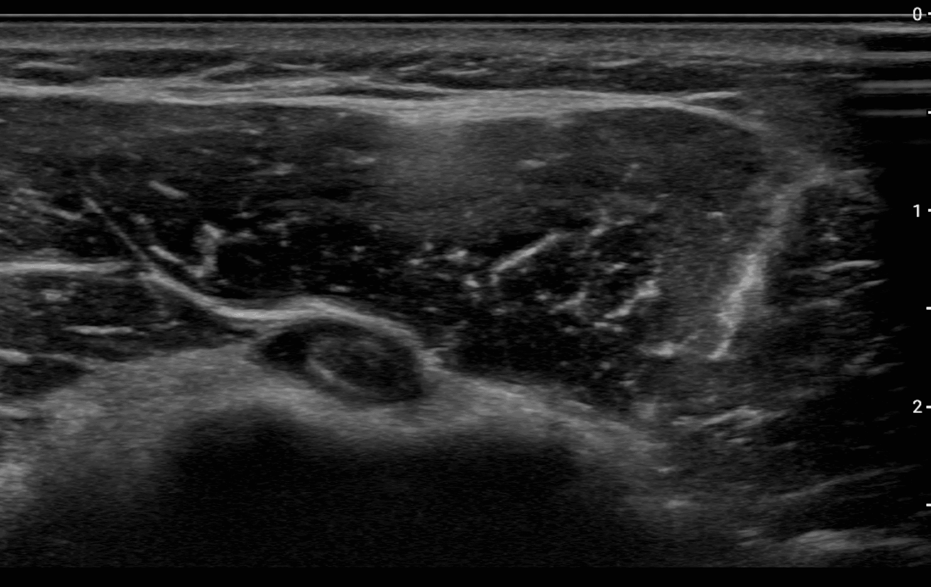
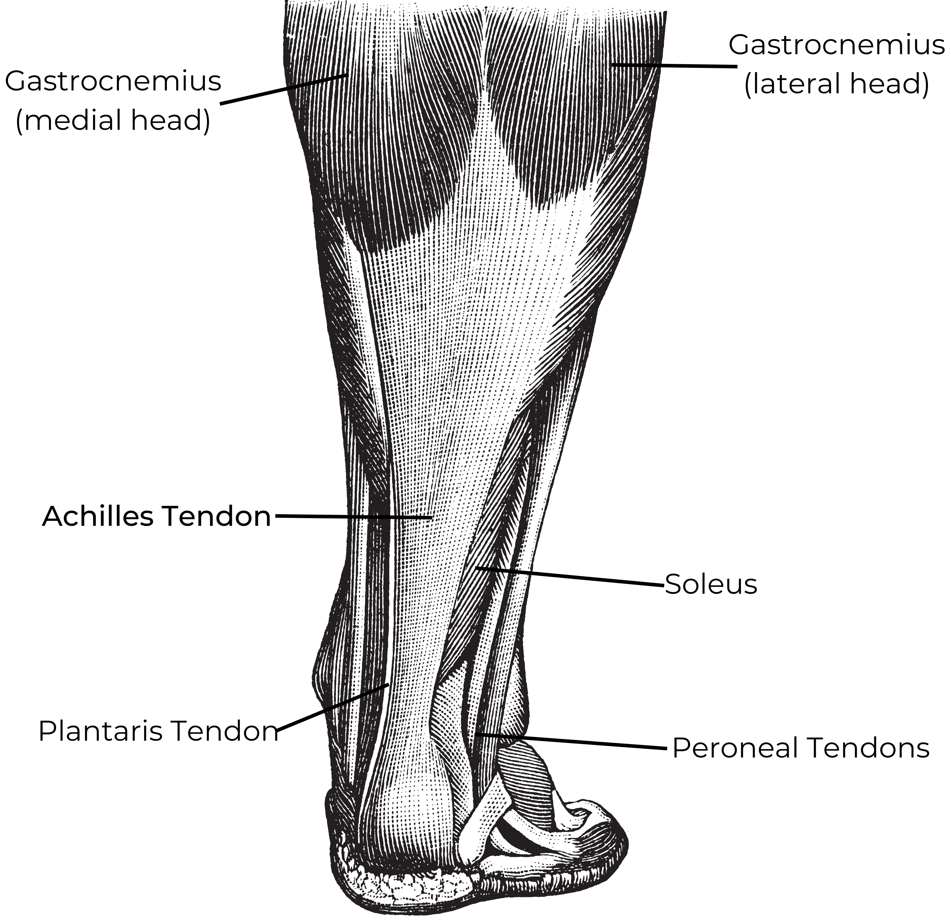
Identify normal features of the Achilles tendon and surrounding anatomy on ultrasound.
Perform specific ultrasound tests that help increase the accuracy of diagnosis of full-thickness and partial-thickness Achilles tendon injuries versus tendinosis.
Target Audience
Physicians, sonographers, and others who perform and/or interpret ultrasound.
Faculty & Disclosure
Faculty
Jason Cox, MD
Program Director, Attending Musculoskeletal Radiologist
Institute for Advanced Medical Education
St. Louis, MO 63141
Disclosure
In compliance with the Essentials and Standards of the ACCME, the author of this CME tutorial is required to disclose any significant financial or other relationships they may have with commercial interests. Jason Cox, MD discloses no such relationships exist. IAME has assessed conflict of interest with its faculty, authors, editors, and any individuals who were in a position to control the content of this CME activity. Any identified relevant conflicts of interest have been mitigated. IAME's planners, content reviewers, and editorial staff disclose no relationships with ineligible entities.
In compliance with the Essentials and Standards of the ACCME, the author of this CME tutorial is required to disclose any significant financial or other relationships they may have with commercial interests.
IAME has assessed conflict of interest with its faculty, authors, editors, and any individuals who were in a position to control the content of this CME activity. Any identified relevant conflicts of interest have been mitigated. IAME's planners, content reviewers, and editorial staff disclose no relationships with ineligible entities.
Credits
* AMA PRA Category 1™ credits are used by physicians and other groups like PAs and certain nurses. Category 1 credits are accepted by the ARDMS, CCI, ACCME, and Sonography Canada.
Course Details
Accreditation
The Institute for Advanced Medical Education is accredited by the Accreditation Council for Continuing Medical Education (ACCME) to provide continuing medical education for physicians.
The Institute for Advanced Medical Education designates this enduring material for a maximum of 1 AMA PRA Category 1 Credit™. Physicians should only claim credit commensurate with the extent of their participation in the activity.
Sonographers: These credits are accepted by the American Registry for Diagnostic Medical Sonography (ARDMS), Sonography Canada, Cardiovascular Credentialing International (CCI), and most other organizations.